Myelogram
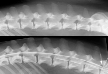
A myelogram is an x-ray study in which special dye is injected into the spinal fluid surrounding the spinal cord. The spinal cord is not visible on a normal x-ray. Injection of this dye outlines the spinal cord, and makes it visible on the the x-ray. The injection of this dye into the spinal fluid may be done in the neck area (cisternal myelogram) or in the lower back area (lumbar myelogram). Just before the dye is injected, a sample of spinal fluid is collected from the patient and is submitted to the laboratory for analysis. A myelogram is a difficult and very delicate diagnostic procedure, and ideally should be done by a veterinary specialist. General anesthesia is required for the procedure.
A myelogram is a diagnostic procedure indicated when a patient has signs of a spinal cord problem such as difficulty walking, or neck or back pain. While a myelogram is an essential diagnostic step in determining the cause of a spinal cord problem, there are some risks associated with this diagnostic test. Myelograms must be done with the patient anesthetized. As with any general anesthetic, there is a very small risk of anesthetic complications, including patient death. At the Veterinary Medical Teaching Hospital (VMTH), the anesthesiology service anesthetizes the neurology patients, and thus the risk of anesthetic complications is low. For several days after a myelogram, some animals may have more trouble walking, particularly if they had difficulty walking prior to the myelogram. This usually resolves in a few days, but in rare cases it may be permanent. Some animals, particularly large dogs may seizure while recovering from a myelogram. The risk of seizures only lasts for 24 hours after a myelogram. Very rare complications of a myelogram include infection, allergic reactions, and injury to the spinal cord. At the VMTH we are experienced in doing myelograms. They are done on a daily basis and serious complications are rare.
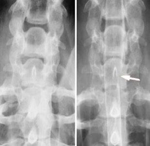
Radiograph (x-ray) of a dog's neck and part of a myelogram. The dog is lying on its back, with the head at the top of the image. The image on the left is a plain radiograph of the neck. The image on the right is part of the myelogram in the same area of the neck. The white arrow is pointing to a disc herniation compressing the spinal cord, only visible on the myelogram. The dog recovered with surgery.