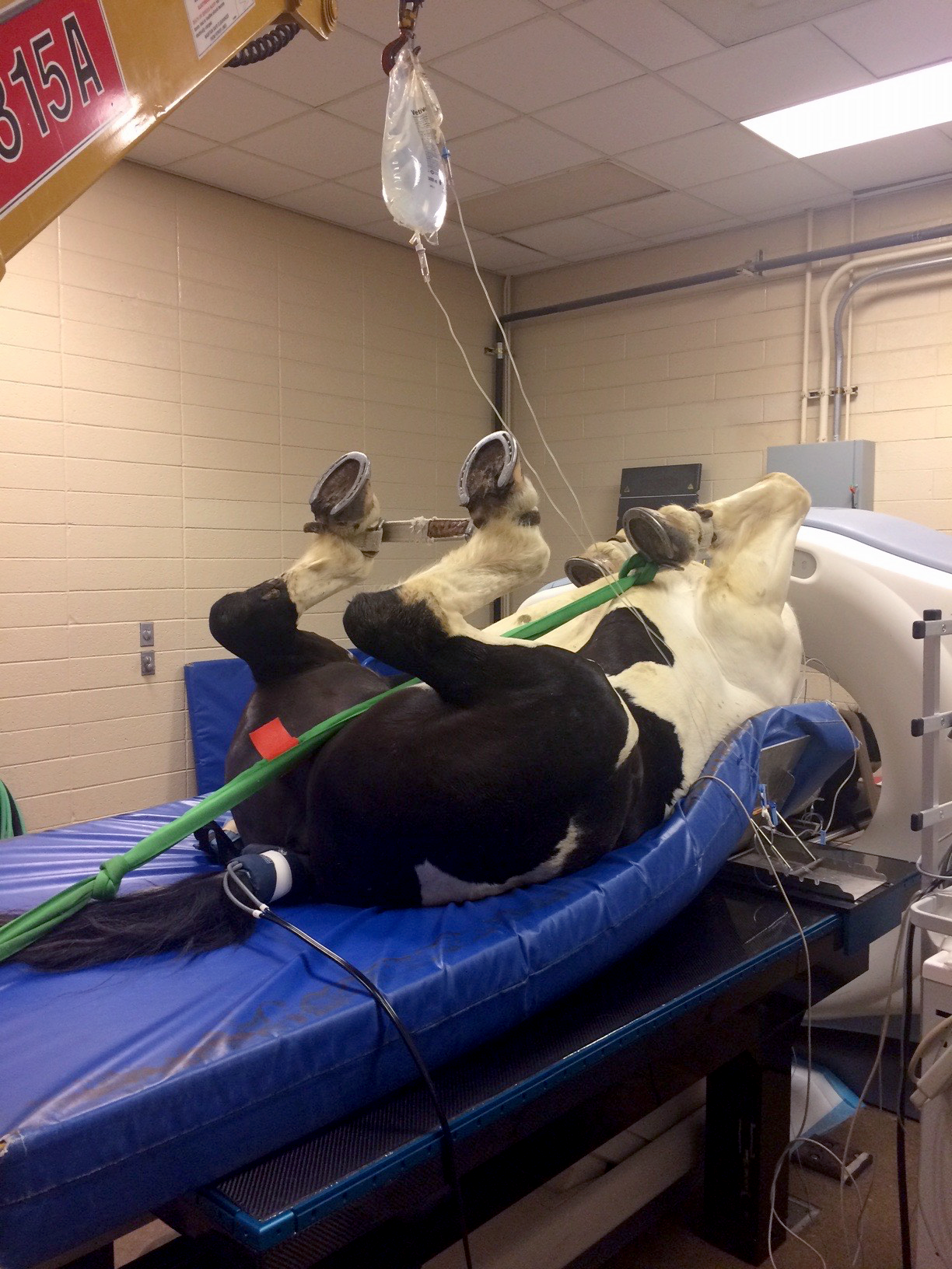
Computed Tomography (CT)

Computed tomography (CT) uses x-rays to produce multiple images of the inside of the body, and provides thin, cross-sectional “slices” for viewing. CT scans of internal organs, bone, soft tissue and blood vessels provide more detail than conventional x-rays. Radiologists use this specialized equipment and knowledge to diagnose problems such as cancer, abnormalities of blood vessels, trauma, and musculoskeletal disorders. CT imaging allows for complete visualization of complex bone structures such as: the carpus, tarsus, and skull. Individual structures can be evaluated in any plane without superimposition of overlying tissues, which allows for better assessment of injuries, and facilitates treatment planning.
Computed tomography is useful for evaluating areas with complex anatomy that are challenging to assess with radiographs and ultrasound. Although commonly the method of choice for bone, CT can also evaluate soft tissue structures. It is very useful to evaluate the equine head, particularly structures such as teeth, sinuses, hyoid apparatus, and tongue. It can be used to identify invasive masses, displaced teeth, and conditions of the eyes and ears. In comparison to other imaging modalities, CT is fairly rapid; a full head scan only takes a couple of minutes.
