Large Animal Ultrasound
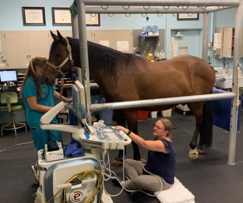
Welcome to the Large Animal Ultrasound Unit at the UC Davis Veterinary Medical Teaching Hospital. UC Davis’ Large Animal Hospital is one of few veterinary hospitals in the country with a service dedicated exclusively to large animal ultrasound and staffed by faculty specifically trained in its use. While the majority of our cases are horses, we also see livestock patients, both production animals and pets, such as goats, sheep and pot belly pigs. We are also called upon to help determine the cause of lameness in everything from an aged Holstein steer to a champion bucking bull. The wide and varied caseload at the VMTH provides our board-certified faculty veterinarians with vast experience and expertise in all areas of ultrasound. As a result, each ultrasound exam can be tailored to the individual needs of your horse to establish the most accurate diagnosis possible. Our state-of-the-art ultrasound machines have been carefully selected for their superior imaging abilities in equine patients. Our mission is to always provide you, your horse and your veterinarian with the highest quality imaging experience, while simultaneously providing an excellent learning opportunity for our students, the equine veterinarians of the future.

Clinical Activities and Procedures
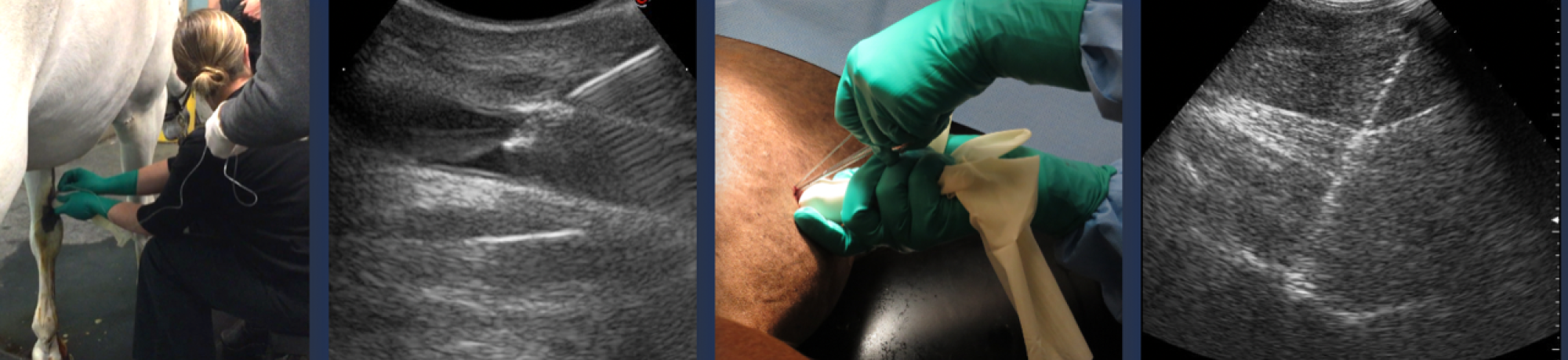
Metacarpal (Fore) & Metatarsal (Hind) Ultrasound Evaluation of the metacarpus or metatarsus is indicated in horses whose lameness has been localized to either region through the use of diagnostic nerve blocks or in horses with regional swelling. Structures evaluated include the superficial digital flexor tendon, deep digital flexor tendon, inferior check ligament and suspensory ligament. Normal tendons and ligaments show an evenly white or echogenic appearance on transverse (cross-section) views and a linear fiber pattern on longitudinal views. Injuries typically cause enlargement (as measured by cross-sectional area) and create hypoechoic (dark) areas within the injured structure as shown in the center image. Ultrasound was used to determine that an inferior check ligament (ICL) injury was the cause of swelling and lameness in this Dutch Warmblood show jumper. The top right image shows severe enlargement of the ICL with a hypoechoic (dark) area and irregular fiber pattern. This can be compared to the normal appearance of the ICL (arrows) shown at bottom right.
The Large Animal Ultrasound Unit offers a complete range of ultrasound imaging procedures. Many studies are performed to examine tendons, ligaments and joints as part of the comprehensive evaluation of horses with lameness or performance problems. Other horses present for thoracic or abdominal ultrasound to evaluate for problems associated with the lungs, heart, abdominal organs or gastrointestinal tract.
Musculoskeletal Examinations include:
- Metacarpal and metatarsal
- Pastern
- Joints
- Neck/Back
- Abdominal
- Cardiac and Thoracic
- Pelvic
- Head
- Ultrasound guided procedures
Metacarpal and metatarsal ultrasound is often performed to diagnose superficial digital flexor tendon or suspensory ligament injuries. Injuries to the deep digital flexor tendon and inferior check ligament do occur but are less commonly identified.
Pastern ultrasound is indicated in horses with lameness localized to the pastern or foot, pastern swelling or digital sheath effusion. Ultrasound can diagnose injuries to the deep digital flexor tendon, distal sesamoidean ligaments, branches of the superficial digital flexor tendon and other small ligaments of the pastern region.
Joint ultrasound examinations of the coffin joint, fetlock, carpus, elbow, shoulder, stifle and tarsus (hock) can be used to diagnose injuries of the collateral ligaments or other regional tendons or ligaments. In horses with wounds, ultrasound can help to determine if the joint or other synovial structure (such as a tendon sheath) has become infected. This information helps to determine the need for additional diagnostic procedures or surgical intervention.
Neck and back ultrasound, such as cervical & lumbosacral ultrasonography can be used in conjunction with radiography to diagnose various conditions such as arthritis of facet joints (cervical & lumbar), sacroiliac ligament injuries and lumbosacral/sacral abnormalities. Transrectal examination of the lumbosacral region is used to detect abnormalities of the lumbar and lumbosacral disc spaces, sacroiliac joint, sacrum and sacral nerve roots.
Abdominal ultrasound is often performed by our service in horses with chronic or recurrent colic, fevers, weight loss, diarrhea, urinary problems or suspect masses. These exams can be very rewarding, often leading to a diagnosis of internal abscessation, liver disease, renal disease, colitis, inflammatory bowel disease, peritonitis and even neoplasia (cancer). In horses with acute colic, abdominal ultrasound is routinely performed by our Equine Surgical Emergency and Critical Care Service to help determine the need for surgical intervention.
Cardiac and thoracic ultrasound is also performed by our service, although many horses with pulmonary or pleural disease are imaged by our Equine Internal Medicine Service. Thoracic ultrasound is primarily indicated in horses with fevers, nasal discharge, abnormal lung sounds or coughing. Although not cardiologists, our ultrasound faculty do have specialized training in large animal cardiology, and also work closely with the hospital’s board-certified cardiologists to properly evaluate any cardiac concerns in horses. Echocardiography is indicated in horses with murmurs, reduced exercise tolerance and other signs of cardiac disease.
Pelvic ultrasound is used for the diagnosis and localization of pelvic fractures is an area of particular interest to the Large Animal Ultrasound Unit. Over the past 10 years, the unit has diagnosed nearly 100 affected horses. Depending on the fracture location and severity, many horses can have a successful outcome after a period of confinement. Although radiography or nuclear scintigraphy (bone scan) can detect pelvic fractures, ultrasound is quick, less expensive and does not require general anesthesia.
Head ultrasound is usually performed in horses with regional swelling to determine its cause. Potential abnormalities include abscessation due to tongue foreign bodies, temporomandibular joint infection, salivary abnormalities (abscessation or sialoliths), osteomyelitis (bone infection) or fracture of the mandible or skull.
Ultrasound guided procedures are performed on a daily basis by the unit. Ultrasound guidance ensures that an intralesional product (such as stem cells, platelet rich plasma, etc.) is placed precisely within the injured portion of a tendon or ligament. It also ensures accurate placement of therapeutic agents into joints, tendon sheaths or bursae, including the cervical facets, sacroiliac joint, hip joint, shoulder joint and bicipital bursa. Ultrasound guidance is also routinely used to obtain samples from abdominal organs (liver, spleen, kidneys), masses or abscesses in horses and other large animal species.
Faculty

Betsy Vaughan, DVM, DACVSMR
Professor
Section Head - Large Animal Ultrasound Unit

Charlène Pigé, DVM, DACVR-EDI
Assistant Professor

Suzanne Brenner, DVM
Staff Veterinarian

Mary Beth Whitcomb, DVM, MBA, DECVDI (LA - Associate)
Professor Emeritus
House Officers

Angela Fleming, BEqSc, BVSc
Fellow - Large Animal Ultrasound
Staff

Rachel Eya
Technician
Frequently Asked Questions
- How do I schedule an ultrasound appointment?
- The Large Animal Ultrasound Service does not function as a primary care service, therefore, animals generally undergo an ultrasound exam after being evaluated by a primary care service, such as the Equine Surgery and Lameness Service, Equine Medicine Service, or Livestock Medicine and Surgery Service. Specific ultrasound appointments for such cases are not generally made, as it is difficult to predict when a lameness evaluation will be completed, especially in horses with complex or multiple limb lameness. In general, cases are seen on a first come, first serve basis throughout the day, although outpatients, recheck ultrasound & emergency exams are given priority.
- What if my veterinarian has already worked up my horse and I just need an ultrasound exam?
- Your horse may qualify for an ultrasound consultation appointment which are available on a limited basis. These are referral appointments and are scheduled by your veterinarian after he or she has contacted an ultrasound faculty member to determine if a consultation appointment is the best option for your horse. If so, you will be contacted by either your veterinarian or hospital admissions to schedule an appointment. As these appointments are designed solely as a consultation service, a lameness exam or additional diagnostics cannot be performed. If it is anticipated that further diagnostics may be necessary, the appointment should be scheduled through our Equine Surgery and Lameness Service.
- What will ultrasound show that radiographs do not?
- In general, radiography (x-rays) is used to evaluate bony abnormalities, and ultrasound is used to evaluate soft tissue structures such as tendons and ligaments. Although ultrasound does provide information about the surface of bony structures, it cannot penetrate bone. Many horses require both imaging procedures to adequately evaluate all bone and soft tissue structures in a region of interest.