UC Davis Becomes Only Veterinary Hospital in the World with a Volumetric PET Scanner
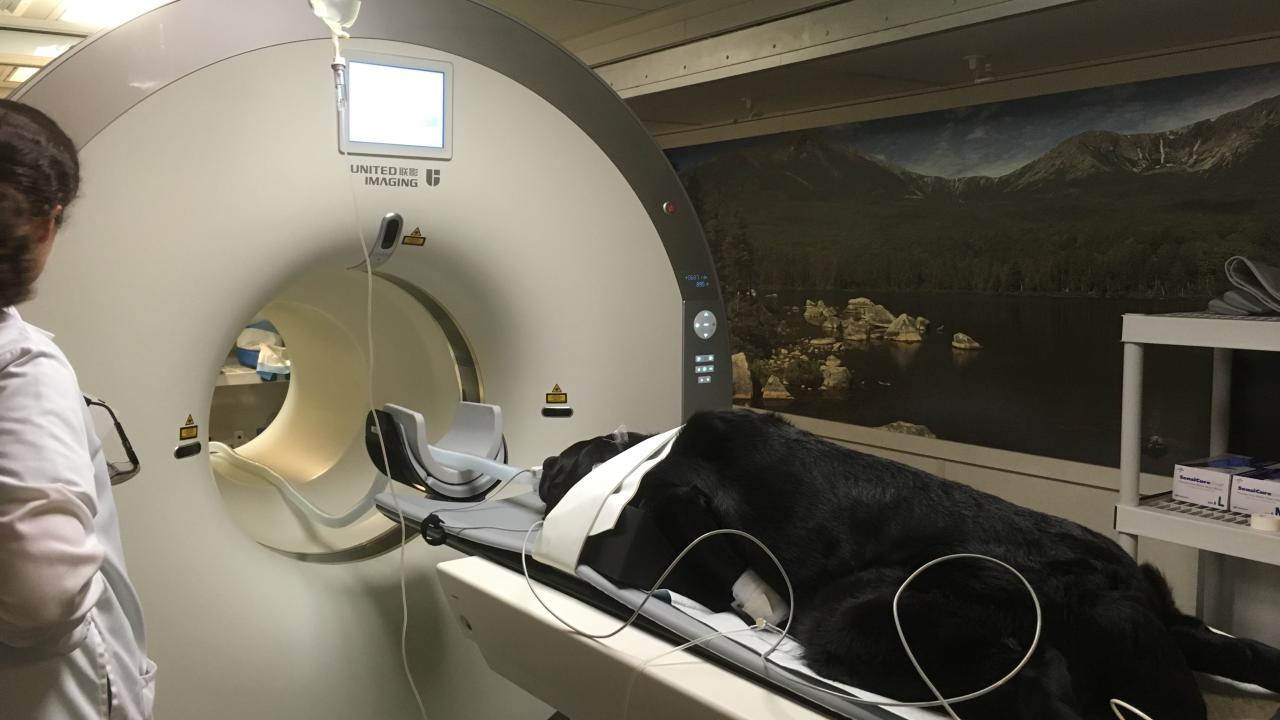
In May, the UC Davis veterinary hospital started imaging patients on a new PET/CT scanner known as the Mini Explorer II. This advanced imaging system, the first of its kind in the world, is designed to provide veterinarians with both anatomic and physiologic information.
“The applications of this machine are far above and beyond typical PET scans,” said Dr. Derek Cissell of the Diagnostic Imaging Service. “We are able to get better spatial resolution and sensitivity that aid in providing the best care possible for our veterinary patients.”
Typical scans can take up to 30-45 minutes to get the information needed to make a proper diagnosis, though radiologists believe that the increased sensitivity of the new scanner may allow them to reduce this time to as little as 5-10 minutes for some types of studies. This is possible because the width of its PET detector is much wider than a conventional scanner, allowing a volumetric acquisition. As an example, the head, neck, and abdomen of a 30-pound dog can be scanned in two acquisitions instead of the multitude of readings that would be required with a conventional scanner.
These faster scans reduce patient anesthesia time, yet still provide outstanding image quality to evaluate disease processes with greater sensitivity and precision.
This may also lead to a reduction of the injectable dose of radiopharmaceutical needed to acquire images. Imaging specialists at UC Davis will be exploring the lowest possible dosage needed to achieve the best image quality.
“That is important in terms of how long we have to confine the patients after completing a scan,” said Dr. Erik Wisner, director of Imaging Services. “If we can get the dosage down to one-tenth of a conventional dose, we can potentially get those patients discharged on the same day.”
One of the most advantageous uses of PET in human medicine is to stage cancer, Wisner explained. While other forms of imaging (MRI, CT, radiographs, and ultrasound) are all representative of imaging anatomy—imaging things you can touch and see—nuclear medicine instead maps out physiologic or metabolic processes.
“Cancer cells behave differently than normal cells in terms of glucose metabolism,” said Wisner. “The radiopharmaceutical agent used in PET scanning behaves metabolically just like glucose, so it is taken up by cells. When one of those cells is cancerous, the agent becomes trapped in the cell, and it cannot be metabolized. A metastatic lesion in a lymph node might not be noticeable on a CT, but the agent trapped in the cell stands out on a PET scan.”
In addition to cancer applications, the PET scanner will also be used for orthopedic, neurology, and cardiology diagnosis, among others.
“This PET scanner has a clear potential to boost our understanding of complex problems,” said Dr. Denis Marcellin-Little of the Orthopedic Surgery Service. “It is providing novel information that will help us detect the sources of pain in abnormal bones and joints, and has clear advantages over other imaging modalities.”
“The design is truly revolutionary,” said Wisner. “[Department of Biomedical Engineering faculty member] Simon Cherry designed this and its prototype. The volumetric acquisition greatly increases the speed and allows us to do a lot of things you could not do with a conventional PET scanner.”
Cherry collaborated with Ramsey Badawi, a physicist in the Department of Radiology at UC Davis Health, to create Mini Explorer. Their end goal is to build a full human-size PET scanner, larger than any currently available. But they needed to start with a scalable prototype first.
In consultation with Drs. Cissell, Wisner, Allison Zwingenberger, and Mathieu Spriet, the team built Mini Explorer I for testing at the veterinary hospital for approximately five months before it was moved to UC Davis’ California National Primate Research Center. They followed with the current Mini Explorer II – a slightly larger model designed to fit a medium-sized dog and other small animal patients.
A few other veterinary hospitals in the country are utilizing PET, but with this new technology, UC Davis is currently the only medical institution in the world with a volumetric PET scanner. The human-sized PET scanner Cherry and Badawi envision will be available for use at UC Davis Health by 2019.
With the UC Davis veterinary hospital performing nearly 1,200 CT scans per year, this additional machine will also be available to take some conventional CT spillover and allow the imaging team to utilize additional CT scanning timeslots on the new machine. This will certainly prove valuable in unscheduled emergency situations that otherwise would cause delays in the regular CT schedule.
# # #