Contact
Equine Surgery and Lameness Service
Telephone
(530) 752-0290
Location
UC Davis Health Science District
VMTH
1 Garrod Drive
Davis, California
Equine Surgery and Lameness Service
Welcome to the Equine Surgery and Lameness Service at the UC Davis Veterinary Medical Teaching Hospital. We offer a wide range of diagnostic, surgical, and therapeutic options for horses, including lameness evaluation, soft tissue and orthopedic surgical procedures, and advanced imaging techniques.
Our surgical faculty and resident veterinarians carefully evaluate each patient before providing a diagnosis and plan for treating the problem. If surgery is deemed necessary, we provide the most advanced surgical techniques available, including minimally invasive surgical techniques such as arthroscopy, laparoscopy, thoracoscopy, endoscopic laser surgery and urinary tract lithotripsy. Additionally, the service offers a variety of regenerative medicine modalities (autologous conditioned serum, platelet rich plasma and stem cells) for treating soft tissue and orthopedic injuries, and joint disease.
In many instances, case management is optimized through consultation with the respective complimentary specialty services available throughout the hospital to provide the most comprehensive care for all surgical disorders. Several conditions are managed through joint efforts by multiple services including neurology, oncology, dentistry and sinus conditions, upper and lower respiratory disorders, and conditions involving the skin, abdominal, thoracic, and reproductive tracts.
Decisions regarding diagnosis and treatment are always made with the goal of optimizing patient outcome.
Clinical Activities and Procedures
Lameness Evaluation
Faculty and resident veterinarians with the Equine Surgery and Lameness Service perform comprehensive evaluations on horses of all breeds and disciplines with a variety of lameness or performance problems. Lameness evaluations include a detailed physical examination and careful observation of the horse in motion. Following static and dynamic exams, local anesthetic techniques are typically used to localize the source of lameness. Once the lameness is localized, imaging techniques such as radiographs (x-rays), ultrasound, computed tomography (CT scan), magnetic resonance imaging (MRI), or positron emission tomography (PET scan) may be used to definitively diagnose the underlying problem and determine the best therapeutic approaches to manage and treat the lameness. More elusive lameness problems may benefit from broader imaging techniques such as nuclear scintigraphy to narrow down their origin.
Minimally Invasive Surgery
Minimally invasive surgical techniques (sometimes referred to as keyhole surgery) are used wherever possible to decrease recovery time, improve healing, and minimize complications. The use of small cameras and specialized instruments introduced into body cavities allow detailed examination and treatment of internal structures through small portals. Arthroscopy and tenoscopy are used to evaluate joints and tendon sheaths, remove bone chips, and address injured cartilage and soft tissue structures. Laparoscopy is used to diagnose and treat problems in the abdominal cavity and can include removal of ovaries or retained testicles, evaluation of chronic colic, and biopsy of various abdominal organs. Endoscopic surgery is frequently used to treat conditions of the upper respiratory tract, reproductive tract (removal of uterine cysts), and urinary tract (removal of bladder stones).
Regenerative Medicine
A majority of our equine patients that present for lameness disorders are treated with regenerative medicine techniques, which include autologous conditioned serum, platelet rich plasma, and stem cells. These therapies are aimed at breaking the inflammatory cycle through neutralization of inflammatory mediators and delivering high concentrations of the cells and cytokines necessary to promote optimal healing of injured tissues .
Advanced Fracture Repair
The Equine Surgery and Lameness Service offers treatment for simple and complex fractures for horses, as well as all of the large animal patients presented to the different services throughout the hospital. We provide the most advanced diagnostic imaging and orthopedic techniques available in our approach to fracture repair. We assess the fracture configuration and blood supply using CT scans before selecting the best treatment methods, which routinely include the use of locking compression plates to provide maximal stability to the fracture repair and optimize post operative patient comfort and fracture healing. Following fracture repair, specialized anesthetic and sling recovery techniques are utilized to optimize recovery from anesthesia. We strive to improve the success rate and outcome for horses with these life-threatening injuries.
Sling Support
The Anderson Sling and the UC Davis Large Animal Lift are invaluable pieces of equipment used to reduce the risk of injury during recovery from anesthesia and improve patient outcomes. Although the sling is most often used for horses that have undergone fracture repair, it is often used for horses that need special assistance in standing during recovery from general anesthesia. The Anderson Sling also serves as a post-operative support system for horses with critical orthopedic conditions and those at risk of developing support limb laminitis. The VMTH is equipped with hoists that are strong enough to lift draft horses, with hospital personnel trained in the safe and efficient use of the Anderson Sling and UC Davis Large Animal Lift.
Hospital Care
Patient care at the VMTH is provided 24 hours/day by trained and dedicated personnel. Our facilities include a 40-stall barn, a 10-stall intensive care unit (ICU), a neonatal ICU, and an isolated infectious disease ICU. We follow stringent procedures regarding infectious disease control to prevent the spread of infection. We also require ongoing surveillance of all hospitalized patients to minimize the risk of transmissible infections.
Other Specialties
Other procedures offered include the diagnosis of upper respiratory conditions utilizing overgound exercising endoscopy, sinus disease and dental abnormalities, surgical oncology, reconstructive surgery, laser surgery, and performance evaluation of equine athletes.
All Species Imaging
The UC Davis All Species Imaging Center is equipped with a Qalibra CT scanner which has the largest gantry available at 90cm. The scanner is capable of imaging heads and distal limbs in the standing sedated patient, and the entire cervical spine (including CT/myelogram), stifles, and pelvis in horses under anesthesia. All Species Imaging offers both low field and high field MRI capabilities for horses. Low field MRI is useful for imaging the distal limb in the standing sedated patient, while the 3 Tesla magnet of the high field MRI produces unparalleled image quality and can image heads and limbs from the proximal cannon bone to the foot in the anesthetized patient. Nuclear medicine techniques offered include scintigraphy and PET scanning. Scintigraphy can be used to image large regions (front end, hind end, or whole body) for increased skeletal metabolic activity, which often corelates with areas of injury. PET scanning provides a more focused examination of the skeletal, soft tissues, or both in a specific region to identify areas of increased metabolic activity which frequently correlate with areas of injury. PET scanning is frequently used in conjunction with CT scanning or MRI to provide the most comprehensive diagnostic imaging for our equine patients.
Faculty
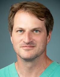
Scott Katzman, DVM, DACVS
Associate Professor
Chief of Service
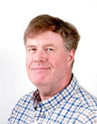
Carter Judy, DVM, DACVS
Clinical Professor
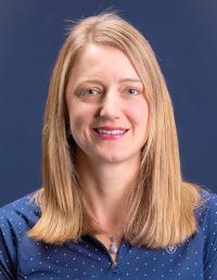
Heidi Reesink, VMD, PhD, DACVS
Professor
House Officers
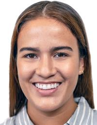
Stephanie Ortiz Gutierrez, DVM
Resident III
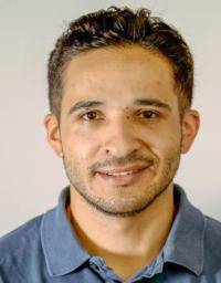
David Orozco Lopez, DVM
Resident II
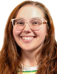
Amber Best, BVMS
Resident I
Staff
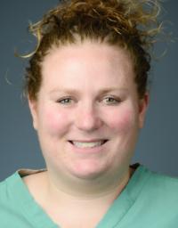
Sarah Blasczynski, BS, RVT, VTS
Service Manager
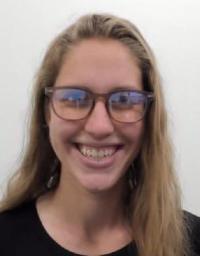
Abigail Jensen
Equine Surgery Coordinator
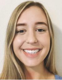
Jessica Behar
Animal Health Technician
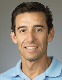
Gabe Gil
Animal Health Technician
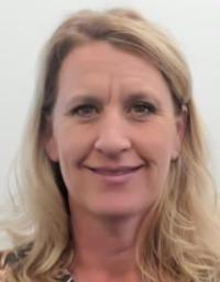
Paige Allenbaugh
Large Animal Surgery Technician
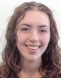
Rachel Herstik
Large Animal Surgery Technician
