Advances in Diagnostic Imaging Improve How Radiologists “See” Horses’ Legs
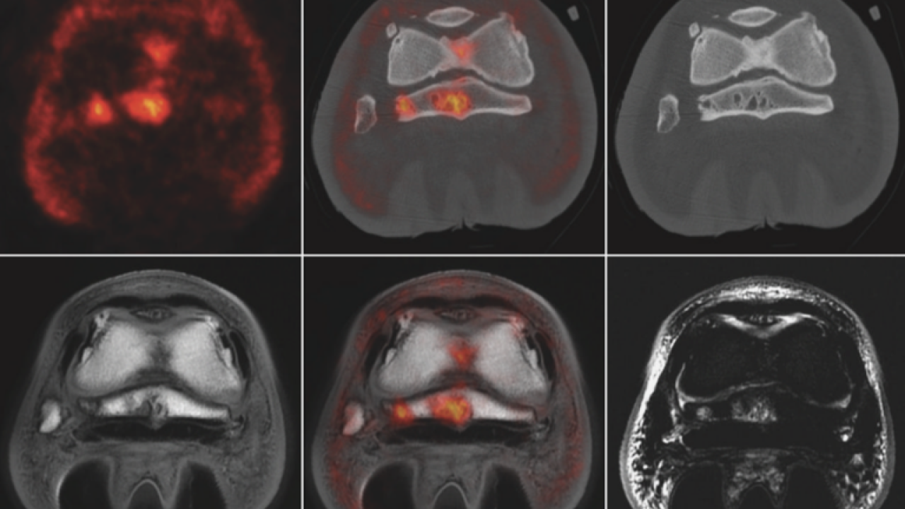
Scientific journals Veterinary Radiology & Ultrasound, Equine Veterinary Education, and the Equine Veterinary Journal are celebrating the publication of nearly 100 equine imaging papers in the last five years. In particular, the evolution of computed tomography (CT), the introduction of positron emission tomography (PET), and the continued growth of magnetic resonance imaging (MRI) have transformed how we image horses’ legs and have led to important clinical advances. UC Davis radiology professor Dr. Mathieu Spriet, who pioneered the use of PET in horses, was one of the guest editors for this special issue.
The virtual collection of 20 papers—selected by Drs. Spriet, Ann Carstens, and Tim Mair—covers advances in CT, PET, MRI, ultrasound, radiography (x-rays), and nuclear scintigraphy. It highlights new designs, advances in robotics, novel approaches to imaging challenges, and the role of comparative imaging studies.
“We live in a fascinating time for equine imaging,” said Spriet. “In the last 20 years or so, we went from relying mainly on x-rays and ultrasound, to now having a diverse arsenal of imaging techniques. The better understanding of musculoskeletal injuries and the ability to identify them earlier provide tremendous opportunities to improve horse welfare and safety.”
One of the most significant advances in equine imaging is the ability to perform these diagnostics on standing sedated horses. Previously, imaging required horses to undergo general anesthesia, which adds to the cost, requires additional staff and equipment, and in rare cases, results in adverse reactions to anesthesia drugs. The ability to utilize these technologies on standing horses under sedation allows for more routine use and provides more options for patients that are not able to undergo anesthesia. In the case of PET, it has also led to use as a veterinary screening tool at Thoroughbred racetracks, which has contributed to reduced injuries and fatalities.
Now in its third year of application at Santa Anita Park, PET scans have benefited more than 500 horses at the renowned racing facility. With the rapid expansion in the availability of this technology - a total of 10 different sites are expected to be equipped with the technology by the end of 2023 – PET is becoming accessible to more diverse populations of horses. Standing PET can be used from the foot to the knee in the front limbs and from the foot to the hock in the hind limbs.
These exciting new imaging modalities have revolutionized how we diagnose lameness in horses. Just imagine what the next five years will bring!
The virtual issue is free for 12 weeks and can be found here.
# # #