Innovation
Veterinary dental surgeons and a UC Davis expert in the biomechanics of cartilage and cartilage healing processes adapted cutting-edge biomedical technology to regrow jawbones in dogs that lost bone to injuries or removal of cancerous tumors. The team used a titanium plate, a scaffolding material and a bone growth promoter to achieve successful outcomes in more than 20 dogs.
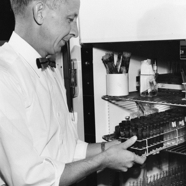
Also at the teaching hospital, the Nutrition Support Service developed new ways to analyze amino acids and other ingredients in pet food and formulate appropriate diets to aid in treatment of hospital patients.
Known for decades for equine research, the school faculty introduced the "track in a box" racetrack surface simulator to assess how different surfaces affect the risk of injury on the track. Video analyses of horses' gaits and lameness issues help the racing industry assure the welfare of equine athletes by reducing the risks of catastrophic injury on the track.
In the 1990s, veterinary neurologists, pathologists and radiologists demonstrated the first CT-guided, stereotactic brain biopsies, a novel way to diagnose canine brain tumors accurately and safely. The stereotactic device is a frame that assures stability of the patient during the delicate procedure.
Emergency and critical care clinicians customized airway management systems, advanced fluid therapy, intravenous catheter care protocols and CPR standards for small animals.
Equine faculty co-invented the Anderson Sling and Large Animal Lift to support horses safely during recovery from injury or surgery; faculty also adapted the sling for use in airlift rescues of large animals. Earlier specialized mechanical equipment was developed such as adjustable tables for horses and other outsized animals. Imaging specialists crafted customized instruments to perform MRIs and computed tomography scans on animals as small as a cat and as large as a horse. Veterinary radiologists also were the first to describe a scintigraphy technique that became the norm for evaluation of a serious defect of the circulatory system known as a portosystemic shunt.
At the Center for Companion Animal Health, the linear accelerator and built-in CT scanner, the most advanced instrument of its kind, allows veterinary oncologists to pinpoint tumors and assure precise doses of radiation therapy to cancers in large and small animals. The equipment senses motion so that clinicians can stop treatment if a patient moves even slightly out of range. Facilities accommodate large animals, protect staff from radiation exposure and provide a dedicated recovery area for equines.
Advanced computer technology permits epidemiologists to analyze large data sets, sort records for retrospective clinical research studies and design complex models of disease for large-scale research projects.
Toxoplasma gondii is a one-celled parasite responsible for the deaths of many endangered sea otters living off the California coast. Radio tagging, telescopes, and other tools, along with the expertise of pathologists and parasitologists, have allowed faculty to detect, isolate, and unravel the life history of parasites in sea otters. Using these tools, scholars found that the biggest risk to the animals comes from domestic cat waste that runs off into the ocean. This work helped establish sea otters as a sentinel species for marine ecosystem health. In addition to posing a danger to animals and the environment, toxoplasmosis can occur in pregnant women.
To optimize physical rehabilitation, underwater treadmills and other equipment are adapted especially to fit small animals. Faculty are also among the few who can design customized orthotic devices for their patients.
The equine treadmill at the Equine Athletic Performance Laboratory faculty has served as a powerful tool for assessing lameness or other performance issues in horses and in conducting research on equine athletes.
Technology aided in reducing the number of animals used in teaching. Plastination to preserve reusable specimens and software courses reduced animal use in the curriculum while maintaining high quality instruction. State-of-the-art surgical laboratories, supervised by faculty and technicians, now allow students to perform simple procedures that benefit animals and improve their welfare.
Several "core facilities" employ technology and veterinary expertise to provide up-to-the-minute research support, collaboration and educational opportunities: the Biological Media Services; Comparative Pathology Laboratory; Flow Cytometry - UC Davis School of Medicine; Real-time PCR Research and Diagnostics Core Facility; the Advanced Imaging facility in the Health Sciences District; and the University of California Veterinary Medical Center - San Diego.