
Routine Training at UC Davis’ New Imaging Center Diagnoses Sialolith in Horse’s Cheek
“Case of the Month” – October 2025
Four years ago, Joe Juice, a then 15-year-old American Quarter Horse gelding, had a sialolith removed from the right side of his face at the William R. Pritchard Veterinary Medical Teaching Hospital (VMTH). He recovered quickly and returned to his role as a member of the UC Davis Center for Equine Health’s (CEH) teaching herd.
Sialolithiasis, or the formation of salivary stones, is not common in horses. The hard masses resulting from the condition typically start small, forming around an item such as a small piece of grass that finds its way into the salivary duct, and grow as layers of calcium are added over time (similar to how enteroliths form). As they get larger, they can rub on the insides of the cheeks, causing ulcerations, which can lead to inflammation, infection, and difficulty eating. If saliva production becomes altered, digestion may be affected.
One study reported recurrence of sialoliths after surgical removal in 24% of cases, with the average time to recurrence noted as just under three years.
Fast forward to the summer of 2025, when Joe Juice’s CEH care team discovered a visible bump on his cheek and suspected another sialolith based on his history.
Around this same time, the hospital’s Diagnostic Imaging Service was preparing to open the All Species Imaging Center. Part of that preparation included training sessions imaging various animals on the new equipment in the center. One of those pieces was the large bore equine computed tomography (CT) scanner.
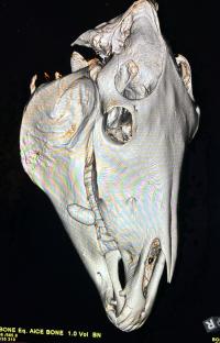
Since Joe Juice was already in need of imaging to confirm the sialolith, his veterinary team decided to utilize him as one of the first training models for the new equine CT scanner. His scan revealed a large, oval, mineral opaque structure measuring approximately 2x3x6 centimeters within the soft tissues of his cheek, consistent with a sialolith.
“Joe Juice was a model patient for our standing CT training,” said Dr. Mathieu Spriet, director of Imaging Services at the VMTH. “His sialolith was definitively diagnosed with the standing CT, giving surgeons a clear and concise assessment of the growth. This equipment’s ability to determine injuries and disease without full anesthesia is already proving to be tremendously valuable for our patients.”
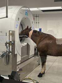
This new equine CT scanner, the first of its kind in California, allows for far more diagnostic capabilities for horses and other large animals. Imaging capabilities using the previous CT scanner were limited to the limbs and head of fully anesthetized horses. This scanner is integrated onto a complex platform that can be moved both vertically and horizontally. That allows head and distal limb scans to be performed on standing horses using only sedation. This technological innovation, combined with the large bore of the scanner, allows for CT imaging of the stifles, pelvis, and entire vertebral column.
Dr. Scott Katzman, chief of the Equine Surgery and Lameness Service, along with equine surgery residents Drs. Stephanie Ortiz and David Orozco-Lopez, removed Joe Juice’s sialolith a few weeks later.
The Anesthesia Service placed Joe Juice under standing sedation, and the surgery team was able to remove the smooth, round-edged mineralized sialolith without complication.
“Joe’s case is a great example of the importance of routine check-ups for your animals,” said Dr. Katzman. “His first sialolith was discovered during a routine procedure several years ago, and this second one was discovered early because his care team routinely examines him for abnormalities. It's also a great example of what we're able to do with sedated standing procedures, instead of full anesthesia.”
Joe Juice was able to return to CEH shortly after surgery and resume his normal physical activity.
“We’re so grateful for the imaging, anesthesia, and surgery expertise at the VMTH,” said Dr. Carrie Finno, director of CEH. “Joe Juice has been a star member of our herd for 17 years. He is one of our go-to horses when it comes to teaching veterinary students. His kindness and patience help them learn about everything from lameness exams to cardiology.”
Learn more about the CEH teaching herd and support them here.
# # #