UC Davis Opens All Species Imaging Center
Donor-Funded Hub of Specialized Imaging Is Most Advanced on West Coast
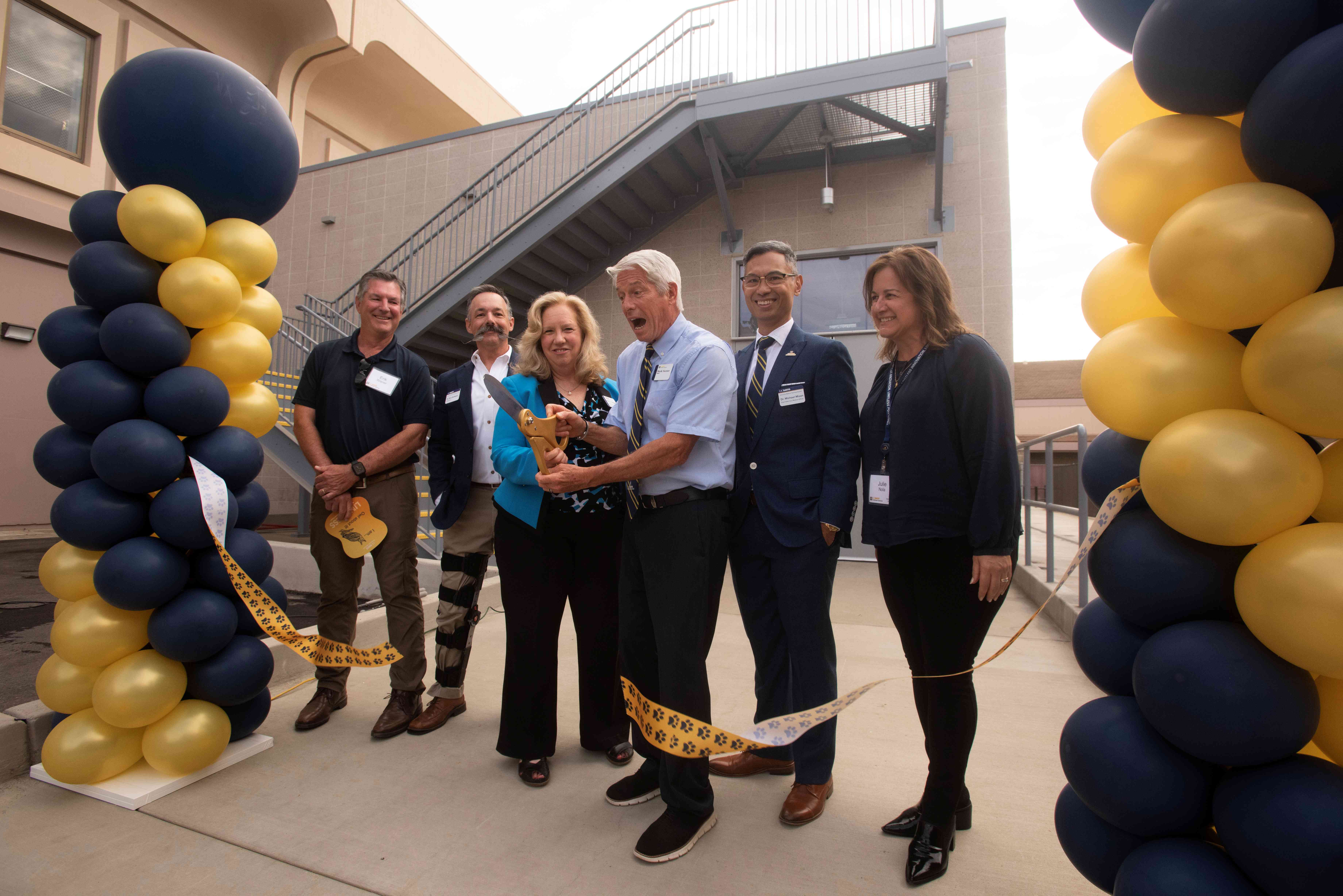
The UC Davis William R. Pritchard Veterinary Medical Teaching Hospital (VMTH) has opened the All Species Imaging Center. This central hub for all advanced diagnostic imaging is the realization of leadership vision and philanthropic endeavors over a decade in the making. Thanks to the generosity of many donors, the entire building and two of the new scanners were donor funded.
The All Species Imaging Center is adjacent to the VMTH and includes four diagnostic imaging suites, two control rooms, a patient holding room, a radiologist’s image interpretation room, and office space for technical staff. The center is outfitted with the latest in advanced imaging technology. The four imaging suites are dedicated to small animal computed tomography (CT), high field magnetic resonance imaging (MRI) for both small and large animals, positron emission tomography (PET)/CT for both small and large animals, and a dedicated large bore equine CT, which is revolutionizing how the hospital diagnoses and treats horses and other large animals.
"With these advanced scanners, we have been able to image areas of animals that we had never been able to scan before, such as the lumbar spine of horses and cows, the pelvis and stifles of horses, a full equine cervical spine, and the abdomen of a llama," said Dr. Mathieu Spriet, director of Imaging Services at the VMTH. "We believe this to be the best veterinary imaging facility on the West Coast and one of the top veterinary imaging facilities in the world."
The new small animal CT scanner and the MRI scanner replaced machines that were in use at the VMTH for more than 15 and 25 years respectively. These state-of-the-art scanners offer significantly higher image quality and much faster image acquisition times. The new imaging technologies also open the door for novel clinical applications such as 4D cardiac imaging, functional brain imaging, and other clinical diagnostic studies that, until now, were not possible. UC Davis is now able to increase diagnostic imaging caseload and fulfill the demand for the highest quality imaging services in California.
The center also serves as home to the world’s largest advanced radiology training program. With 12 residents and eight board-certified radiologists, the hospital’s Diagnostic Imaging Service is the premier training ground for veterinarians seeking board certification in imaging.
The All Species Imaging Center includes:

Small Animal CT Scanner
The new small animal CT scanner improves to a 160 slice scanner from the previous 16 slice scanner that was in use at the VMTH for nearly 20 years. With a detector four times larger (4 centimeters) and rotation speed four times faster (one rotation in 0.25 seconds), scans can be completed orders of magnitude faster, making CT scanning far more efficient than previously. Additionally, the more than 20% wider bore opening (85 centimeters) accommodates easier positioning of patients. Another major advantage of this new CT scanner is in cardiac applications with the faster speed able to reduce motion artifacts created by heart contractions.
High Field MRI
This new MRI, with a 3T (Tesla) magnetic field, replaces the existing 1.5T MRI which was in use for more than 25 years. The new MRI has a 75 centimeter bore – the widest bore available on a 3T magnet. Unlike the previous MRI, this new unit accommodates both small and large animals. For small animals, the MRI is mostly used for neurological cases (brain, spine imaging), orthopedics, and oncology. In large animals, the hospital is now able to image distal limbs and heads, which was not available previously with high-field MRI.

PET/CT Scanner
The new PET/CT scanner is the commercially available version of the prototype used at the VMTH for the past several years. The machine physically combines two scanners, with an 80 slice CT on one side and an extended field of view (45 centimeters) PET on the other side. This combination scanner is mostly used for small animals but can scan some aspects of a horse on the PET side. Most PET scans of horses, however, will continue to be performed on the existing equine PET scanner allowing limb scanning on standing horses. An added bonus of this machine is its capability to be used as an overflow scanner when the patient schedule for the primary small animal CT scanner is at capacity, allowing the hospital to increase its CT volume and more efficiently serve patients.
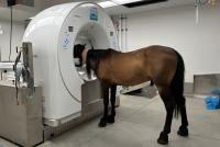
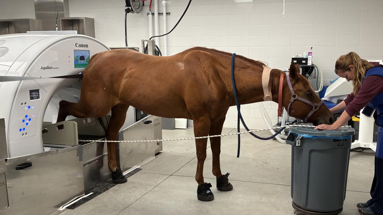
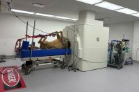
Large Bore Equine CT Scanner
This new equine CT scanner, the first of its kind in California, allows for far more diagnostic capabilities for horses and other large animals. Imaging capabilities using the previous CT scanner were limited to the limbs and head of fully anesthetized horses. This scanner is integrated onto a complex platform that can be moved both vertically and horizontally. That allows head and distal limb scans to be performed on standing horses using only sedation. This technological innovation, combined with the large bore of the scanner, allows for CT imaging of the stifles, pelvis, and entire vertebral column.
# # #