
Surgery Reverses Puppy’s Heart Failure
“Case of the Month” – January 2020
When Ernesto and Chelsea Torres received Riley as a young puppy, they had no idea she had a congenital heart defect. The 4-month-old German shepherd was getting her first immunizations when her veterinarian discovered she had a loud heart murmur. He immediately referred Riley to the Cardiology Service at the UC Davis veterinary hospital.
Once at UC Davis, the full details of the severity of Riley’s heart disease were discovered, and she was scheduled for minimally invasive surgery the next day.
An echocardiogram (ultrasound of the heart) was performed, and Riley was diagnosed with a patent ductus arteriosus (PDA) and mild valve abnormalities. A chest x-ray revealed a severely enlarged heart and evidence of fluid in her lungs – findings consistent with heart failure.
Riley’s condition was the result of a birth defect due to the persistence of the ductus arteriosus, a vascular structure important during development in utero. The ductus arteriosus is a small channel that connects the pulmonary artery (which carries blood to the lungs after birth) and the aorta (which carries blood to the rest of the body). In the womb, it is responsible for conveying blood past the non-functional lungs (since puppies don't breathe air before birth), and into the systemic circulation. At birth, when an animal takes its first breath, the lungs become filled with air. This causes a decreased resistance to blood flow, and blood moves through the blood vessels of the lungs, instead of through the ductus arteriosus. In the normal animal, the ductus arteriosus should fully close after birth. When the ductus arteriosus does not close, extra blood continuously circulates through the lungs and the left side of the heart which can result in heart failure (fluid in the lungs) and death by one year of age.
In Riley, it did not close.
“Riley’s condition was severe,” said cardiology resident Dr. Ashley Sharpe. “She was in heart failure and would likely succumb to her disease within weeks to months had the Torres’ not sought specialty care for her.”
To close the ductus arteriosus, Dr. Sharpe recommended a procedure to stop blood flow through the PDA. This can be achieved by a thoracotomy (open-chest surgery) and manually tying off the ductus, or it can be achieved by a minimally invasive interventional procedure using a catheter to place a small device inside the ductus to stop blood flow. In this method, cardiologists feed a long catheter through the femoral artery and into the aorta (main artery in the body). If corrected, some effects of the animal's PDA are reversible and the prognosis for survival is good, with patients often living a normal lifespan.
“It was scary hearing that she needed heart surgery,” said Chelsea. “Even with the minimally invasive option, there was still a chance that she might not make it. We were very nervous.”
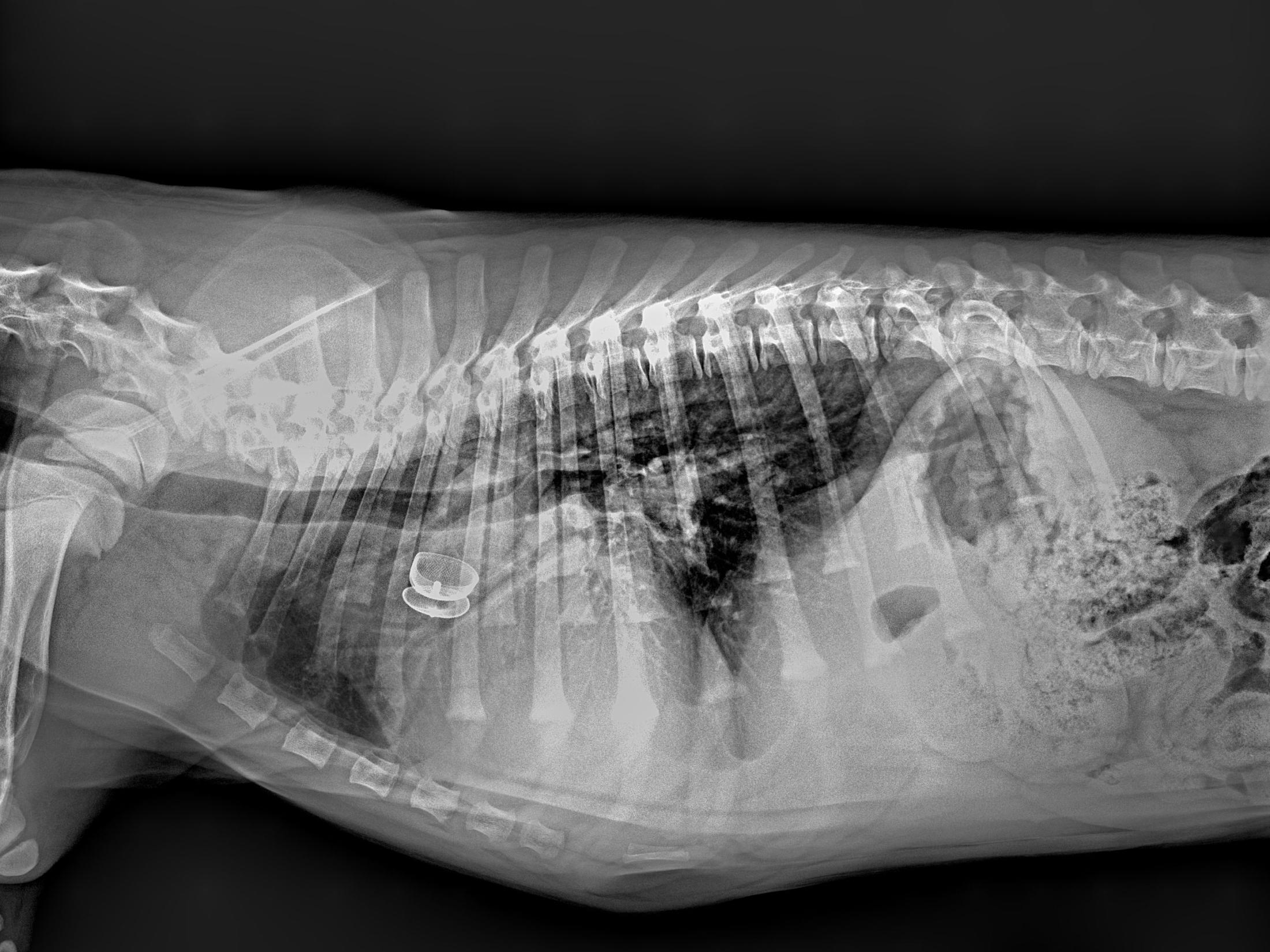
Riley was anesthetized and continuously monitored by the Anesthesia/Critical Patient Care Service. Using fluoroscopic guidance, Drs. Sharpe and Maureen Oldach proceeded with the minimally invasive implantation of an Amplatz Canine Ductal Occluder (ACDO) to occlude the PDA. Surgeons from the Soft Tissue Surgery Service were on hand to assist with a thoracotomy if the cardiologists were not able to complete the implantation via minimally invasive methods. Thankfully, all went well, the ACDO was placed successfully, and Riley recovered from anesthesia uneventfully.
Following surgery, Riley was placed on strict rest to allow the device to stabilize. This has been challenging with a young, energetic puppy that needs to be trained. But Riley has been able to slowly increase her activity level over the past few weeks.
“Based on x-rays and an ultrasound performed at Riley’s recheck appointment, we are happy to report that she is no longer in heart failure,” said Dr. Sharpe. “All of her heart medications have been discontinued, and we anticipate that she will live a normal life.”
“Riley just loved her,” Chelsea said about Dr. Sharpe. “She was wonderful to work with. The entire cardiology team seemed so knowledgeable. She made us feel comfortable that Riley was in good hands.”
Now on her fifth week of recovery, Riley is able to start more activity, which has helped with her training schedule.
“Riley is doing very well,” said Ernesto. “Chelsea and I are very grateful and very impressed with the staff at UC Davis veterinary hospital.”
“We were scared throughout the process,” said Chelsea. “But after what has happened, we feel Riley was placed with us for a reason.”
# # #