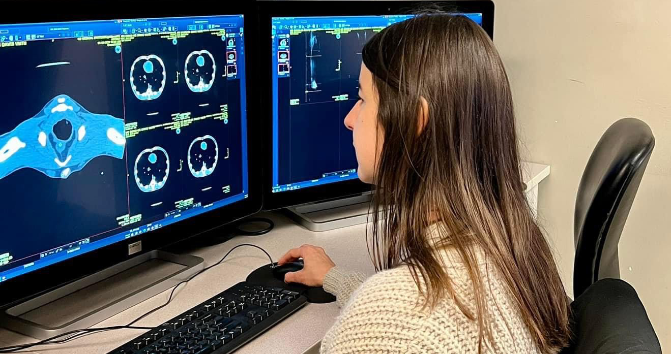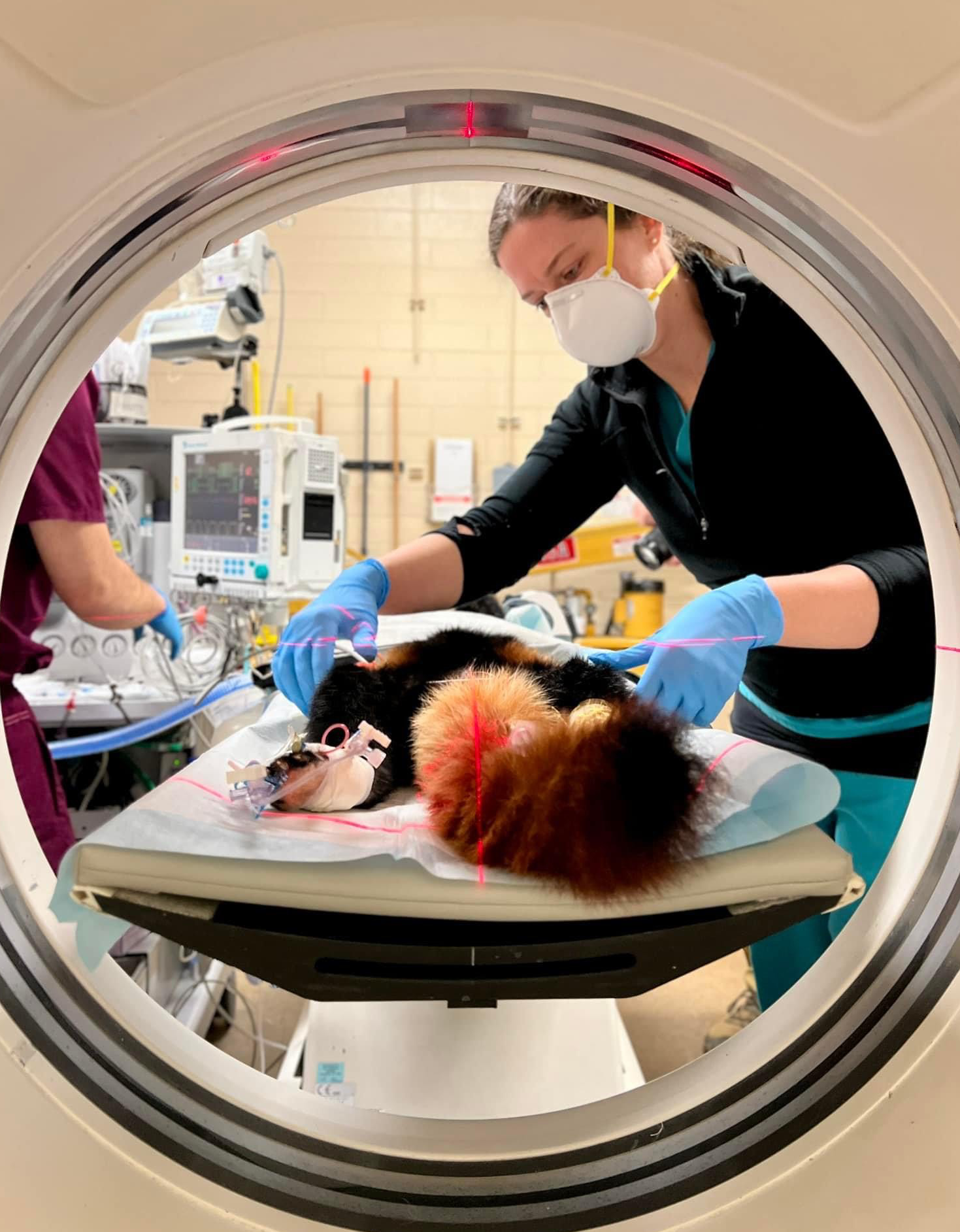
Computed Tomography (CT)
Computed tomography (CT) uses x-rays to produce multiple images of the inside of the body, and provides thin, cross-sectional “slices” for viewing. CT scans of internal organs, bone, soft tissue and blood vessels provide much more detail than conventional x-rays. Radiologists use this specialized equipment and expertise to diagnose problems such as cancer, abnormalities of blood vessels, trauma, and musculoskeletal disorders.
CT imaging is one of the best tools for studying the chest and abdomen because it provides detailed, cross-sectional views of all types of tissue. It is often the preferred method for diagnosing many different cancers, since the image allows a veterinarian to confirm the presence of a tumor and measure its size, precise location and the extent of the tumor's involvement with other nearby tissue. Additionally, it is invaluable for diagnosing and treating spinal problems and injuries to the skeletal structures because it can clearly show small bones as well as surrounding tissues. Veterinary radiologists often use CT to diagnose abnormal blood vessels in the liver, disorders in the abdomen, orthopedic disease, and to plan proper radiation treatment for tumors.