 Magnetic Resonance Imaging (MRI)
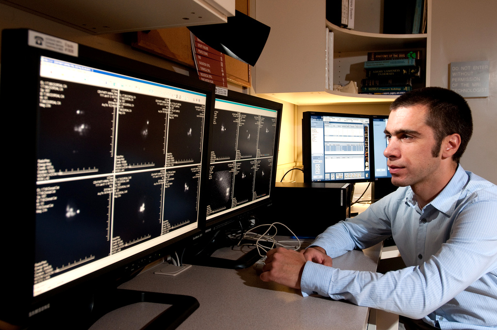
Magnetic resonance imaging (MRI) uses a powerful magnetic field, radio waves and a computer to produce detailed pictures of organs, soft tissues, and other internal body structures. The images can then be examined on a computer monitor or printed. MRI does not use ionizing radiation (x-rays), but detects the motion of protons that are normally present in the body.
Detailed MR images allow veterinary radiologists to better evaluate parts of the body and certain diseases that may not be assessed adequately with other imaging methods such as x-ray, ultrasound or computed tomography (also called CT or “CAT” scanning).
Currently, MRI is the most sensitive imaging test of the head (particularly in the brain) in clinical practice. MRI imaging is performed to help diagnose: tumors and inflammatory diseases of the brain and spinal cord, disorders of the eyes and the inner ear, disease of the pituitary gland, and other problems in the spine.