UC Davis Advances Regenerative Cures
Since 2012, the Veterinary Institute for Regenerative Cures (VIRC) at the UC Davis School of Veterinary Medicine (SVM) has been committed to transforming the SVM into a national leader for veterinary regenerative medicine. VIRC has established laboratory techniques and animal models that have been used to study regenerative therapies for veterinary and human medicine. It has characterized equine, canine and feline stem cells isolated from different tissues with a focus on adult-derived mesenchymal stem cells.
The institute has also developed collaborative, interdisciplinary “disease teams” that include basic research faculty and clinical faculty that focus on “bench to bedside” translation of stem cell therapies. Additionally, VIRC has built strong cross-campus collaborations with faculty in UC Davis’ School of Medicine (SOM), College of Engineering, and College of Agricultural and Environmental Sciences.
Areas of emphasis for VIRC include:
- Manufacturing quality stem cell products for research use and for use in veterinary clinical trials
- Labeling stem cells for in vivo imaging and tracking these cells using state-of-the-art imaging technologies in living patients
- Defining how cells work to heal tissues in in vitro models
- Developing large animal models of diseases to better understand stem cell function in vivo
- Identifying relevant naturally occurring animal models of disease to test stem cell therapy in clinical trials that can benefit veterinary patients and inform human clinical trials
- Training the next generation of undergraduate and professional students, residents and technicians in cutting edge stem cell biotechnology
Led by Dr. Dori Borjesson, VIRC and its collaborators have created several veterinary techniques and procedures that have become, or may soon become, the standard protocol of care in particular cases.
“I have been working on stem cell tracking for about seven years now,” said Dr. Mathieu Spriet, a radiological specialist at the SVM. “My research started with optimizing administration of stem cells to the horse limb. We were looking at different vascular injection techniques, evaluating them with scintigraphy. After trying several techniques, we established that an arterial injection without a tourniquet was the optimal approach. This is now broadly used for front limb injections by many clinicians.”
Significant treatments by VIRC members include:
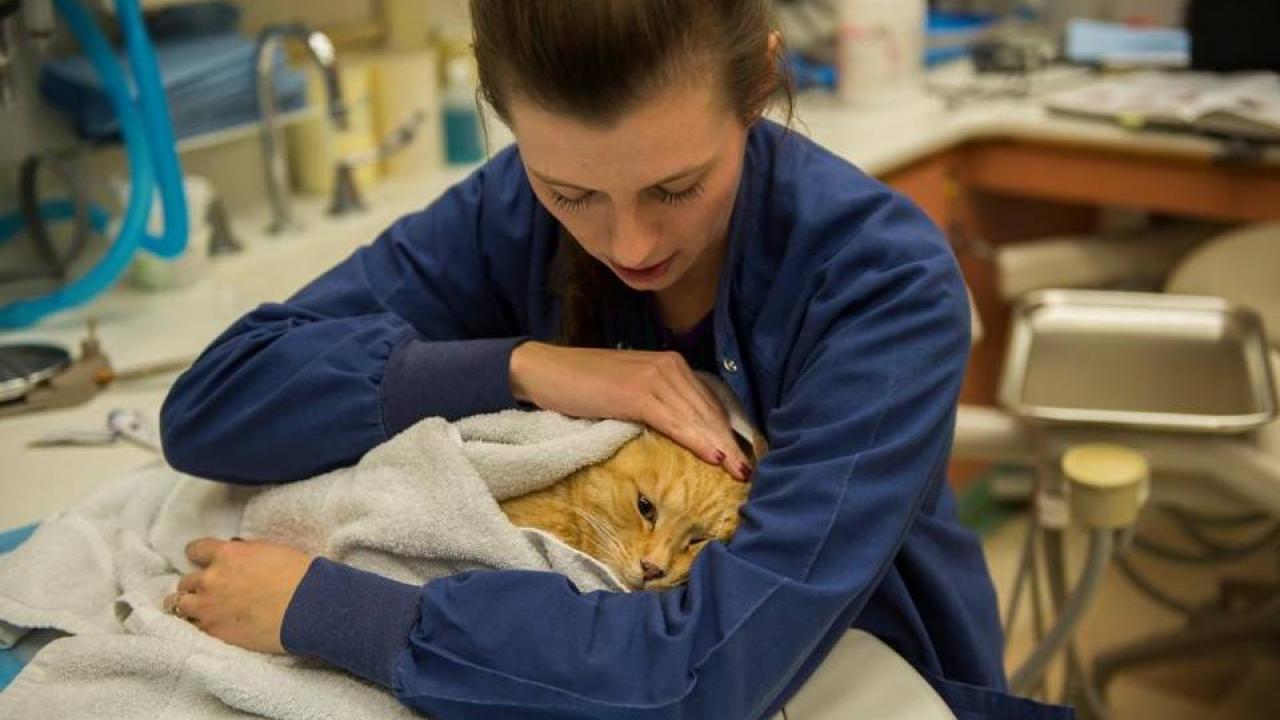
Feline Chronic Gingivostomatitis (FCGS): FCGS is a common, chronic immune-mediated oral disease in cats resulting in severe inflammation and pain. Drs. Borjesson and Boaz Arzi have treated more than 30 affected cats using fat-derived stem cells. Seventy one percent of cats showed significant clinical improvement, and many cats demonstrated complete disease cure. VIRC continues to enroll cats in this study and recently initiated a multi-center clinical trial with Cornell University. Also in development are other clinical studies that involve determining the effectiveness of stem cell therapy in cats during different stages of FCGS.
Spina Bifida: Spina bifida is the most common cause of lifelong childhood paralysis in the United States; approximately four children are born with this devastating defect every day. Spina bifida is the result of incomplete closure of the neural tube which leads to an exposed spinal cord in newborns. Like humans, dogs are naturally affected with spina bifida. Unlike humans, there are no treatment options for these dogs and thus many are euthanized at birth. Drs. Aijun Wang (SOM), Borjesson and Beverly Sturges (SVM) have collaborated to develop a novel stem cell therapy to treat dogs with spina bifida. To date, two dogs have been treated with more being recruited as part of an ongoing clinical trial.
Mandibular Reconstruction: Extensive mandibular defects can be secondary to trauma, tumor resection, or other pathologic, developmental, or congenital disorders. Mandibular reconstruction of critical-size defects requires rigid fixation, typically in the form of a plate and screws, and well-vascularized soft tissues. There are several strategies to fill the critical-size bone defects including autologous bone grafts, bone graft substitutes, and free-fibular flap tissue transfer. These methods, however, are not ideal as they result in donor site morbidity, are limited by graft size, and are difficult to contour. In addition, outcomes may be unpredictable. Drs. Frank Verstraete and Arzi have demonstrated that a regenerative approach to reconstruction of mandibular critical-size defects in dogs using a scaffold and growth factors such as bone morphogenic protein (BMP-2) can be performed successfully and represents an excellent functional solution. To date, they have successfully regenerated the jaws of more than three dozen dogs.
Learn more about VIRC at virc.vetmed.ucdavis.edu/.
This article originally appeared in the August 2018 issue of The Pulse magazine.