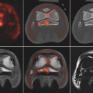
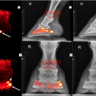
Now in its third year of application at Santa Anita Park, positron emission tomography (PET) scans have benefited more than 500 horses at the renowned racing facility.
UC Davis radiologists were well represented at the 2022 American College of Veterinary Radiology (ACVR) annual scientific meeting, held recently in Reno, Nevada.
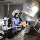
UC Davis’ comparative oncology program, a partnership between UC Davis Comprehensive Cancer Center and Veterinary Medicine that combines human and companion-animal oncology, has been included as part of the renewal of UC Davis’ status as a “comprehensive” cancer center by the National Cancer Institute.
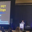
On August 5, UC Davis veterinary radiologist Dr. Mathieu Spriet presented "New Equine Imaging Options with Standing PET at UC Davis," an informative webinar to update veterinarians on the latest in positron emission tomography (PET) for horses.
UC Davis veterinary radiologist and pioneering researcher of equine PET scanning, Dr. Mathieu Spriet was recently selected as one of three veterinary specialists who will review all diagnostic imaging of horses competing in Australia’s 2021 Victorian Spring Racing Carnival.
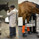
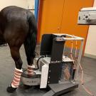
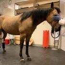
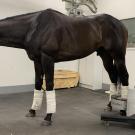
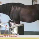
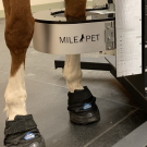
Standing equine positron emission tomography (PET) imaging is not just for racehorses anymore. In the first four months since the installation of the MILEPET scanner at the UC Davis veterinary hospital, 100 horses have been imaged; more than half were performance and pleasure horses.
The UC Davis standing equine positron emission tomography (PET) scanner is officially in use at Golden Gate Fields racetrack in Berkeley, CA, providing imaging at the molecular level to monitor racehorse health and guide training and medical care.
On April 14, 2021, local television morning show "Good Day Sacramento" visited the UC Davis Center for Equine Health. Dr. Mathieu Spriet showcased the latest in equine PET scanning technology.