Dermatopathology and Dermatology
Services
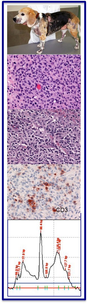
Morphologic features in association with atypical CD3+ T lymphocytes are suggestive of an inflamed T cell lymphoma. Clonality revealed a clonal rearrangement of the T cell receptor of lesions T lymphocytes, confirming the diagnosis of lymphoma. [Click image to enlarge]
- Expert interpretation of skin biopsies
- Consulting and 2nd opinions on previously prepared glass slides
- Clinicopathologic correlations
- Clinical consulting with dermatologist, upon request
- Immunohistochemistry
- Clonality testing for lymphoid lesions
- Reports available via fax or email
Guidelines for Sample Submission
Formalin Fixed samples
- Please ensure that the sample is fully immersed in formalin (appropriate ratio for size of tissue sample to volume of formalin is 1:10).
- If various lesions from different anatomical locations are submitted, collect the sample in separate containers.
- Label container(s) with patient name, sample source, and location.
- Double ziplock-bag the containers and place a paper towel or other absorbent matierial in the first bag, in case of leakage.
Fresh Samples
- If considering sending fresh samples, contact the Pathology office before shipping.
- Fresh samples must be shipped via priority overnight shipping (FedEx overnight express).
- Only ship fresh samples Monday - Thursday, as we do not receive packages on weekends.
- Wrap the tissue sample in saline-soaked surgical gauze (excess fluid should be gently squeezed out). Place tissue with gauze in a tightly sealed container, such as a pill bottle or zip-lock bag, to prevent dessication.
- Place the wrapped sample with some gel cooling packs in a styrofoam box. Ensure that the sample and cooling packs are separated by some paper towels to avoid freezing of sample during shipping.
- Indicate the origin of the tissue sample, especially if they come from multiple sites.
- Ensure the fresh tissue samples are kept separate from formalin-fixed tissues; eg: different bag.
Submission of clinical images
- Hard copies: Submission of clinical pictures is highly appreciated. Add hard copies with the submission form and sample.
- Email attachments: Images can be email to [email protected]. Please clearly state the animal's name, owner's name, and clinic name in your email.
Shipping Instructions
- Complete this electronic Biopsy Submission Form and when you get to the “Mandatory Special Review” section, select “Dermpath”, which will open up a “Veterinary Dermatology specific form”. Please thoroughly complete both of these forms and send a hard copy of the “Derm form” with the wet tissue sample in a second plastic bag.
- Add some padding and absorbent towels before placing specimen in FedEx envelope or priority mail box. Ensure package is crush-proof and will withstand typical shipping conditions.
- Shipping with FedEx: package should be mailed to the address listed below.
- Dermatopatholgy offers a FedEx Shipping Program. To register for an account, fill out and remit a Dermatopathology FedEx Shipping Program form (fillable pdf)
- US mail (USPS): please send packages by priority mail.
Forms
- Biopsy Submission Form
- Dermatopathology Sample Submission form (fillable pdf)
- Dermatopathology FedEx Shipping Program (fillable pdf)
- Dermatology consulting request - In development
Mailing address:
Dermatopathology Service
UC Davis VMTH
One Garrod Drive
VM3A Rm 1346
Davis, CA 95616
Phone: 530-752-1368
Fax: 530-752-7242
Email: [email protected]
Turn around time
- For uncomplicated cases, you can expect a turnaround time of 3 working days after sample arrival.
- Complicated cases requiring additional workup (special stains, immunohistology, clonality) will take longer. You can expect an email from [email protected] with preliminary diagnoses and comments, including an explanation for the delay, within 3 working days.
- For questions regarding these cases, we can be contacted by email at [email protected].
Fees & Billing
- Please contact the Anatomic Pathology office at 530-752-1368 for current fees for Dermatopathology services.
- Accounts: If you already have an account with the William R. Pritchard Veterinary Medical Teaching Hospital, the billing will occur through your existing client account. If you are a new client, a client account will be established upon time of first sample submission.
- Billing: Accounts are invoiced monthly by the William R. Pritchard Veterinary Medical Teaching Hospital and charges are payable upon receipt.
Faculty experts
Dermatopathology
Verena K. Affolter
DVM, PhD, Diplomate ECVP
Professor of Clinical Dermatopathology
School of Veterinary Medicine
Clinson C. Lui
DVM, Diplomate ACVP
Associate Professor of Clinical Dermatopathology
Department of Pathology, Microbiology & Immunology
School of Veterinary Medicine
Dermatology consulting
Catherine A. Outerbridge
DVM, MVSc, Diplomate ACVIM, Diplomate ACVD
Professor of Clinical Dermatology
Department of Medicine and Epidemiology
School of Veterinary Medicine
Professional Links
Events
North America Veterinary Dermatology Forum (NAVDF)
North America Veterinary Dermatology Forum, April 26-29, 2017. Orlando, Florida.
Annual meeting of the International Society of Veterinary Dermatopathology; associated with the NAVDF
Annual meeting of the International Society of Veterinary Dermatopathology; associated with the NAVDF, April 26-29, 2017. Orlando, Florida.
29th Annual Congress of the ESVD and ECVD
29th Annual Congress of the ESVD and ECVD, September 2017. Lausanne, Switzerland.
Societies and Colleges
International Society of Veterinary Dermatology (ISVD)
www.isvd.org
The ISVD is a worldwide association of dermatopathologists and dermatologists dedicated to improve the field of dermatopathology in the veterinary world. Membership is open to veterinarians with an interest in dermatology and dermatopathology. Members have access to post difficult cases and to follow discussion about cases on the ISVD list serve. Annual meetings are organized in conjunction with the North American Veterinary Dermatology meeting and the European Veterinary Dermatology meetings (ESVD/ECVD), the WCVD as well as annual meetings with ECVP/ESVP and ACVP.
American Academy of Veterinary Dermatology (AAVD)
www.aavd.org
The AAVDs mission is to promote scientific progress in veterinary dermatology. Membership is open to veterinarians interested in dermatology. The AAVD is co-organizing the annual NAVDF and the WCVD.
American College of Veterinary Pathology (ACVP)
www.acvp.org
The organization of board certified pathologists promotes excellence in veterinary pathology and oversees specialty training in veterinary pathology. Open to residents and diplomates of this college.
European College of Veterinary Dermatology (ECVD)
www.ecvd.org
The ECVD advances veterinary dermatology in Europe and oversees residency training in veterinary dermatology in Europe. The ECVD co-organizes the annual dermatology meetings in Europe and the WCVD. Open to residents and diplomates of this college.
European Society of Veterinary Dermatology (ESVD)
www.esvd.org
The ESVD is an active international group of veterinarians and scientists interested in biology and diseases of animal skin. The ECVD co-organizes the annual dermatology meetings in Europe and the WCVD. Membership is open to veterinarians interested in dermatology.
European College of Veterinary Pathology (ECVP)
www.ecvpath.org
The ECVPs mission is to advance veterinary pathology and promote high standards within the specialty in Europe. It oversees the residency training in veterinary pathology in Europe. The ECVP co-organizes the annual pathology meetings in Europe. Membership is open to veterinarians interested in dermatology.
European Society of Veterinary Pathology (ESVP)
www.esvp.eu
The ESVP promotes the scientific work of veterinary surgeons and physicians active in the areas of functional and morphologic pathology of animals. The ESVP co-organizes the annual pathology meetings in Europe. Membership is open to veterinarians interested in pathology.