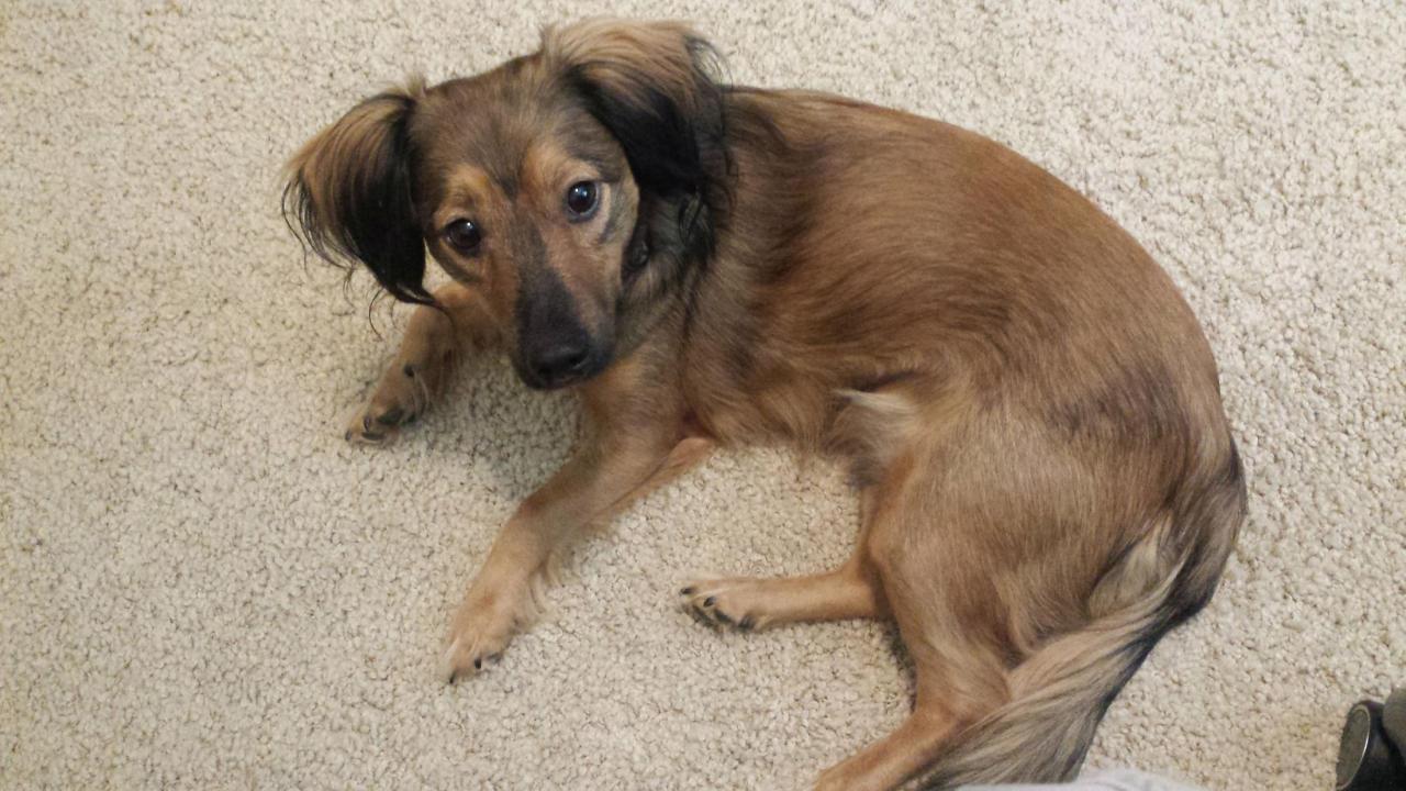
UC Davis Saves Dog’s Eye after Stick Pokes Socket
"Case of the Month" – September 2017
Arrow, a 2-year-old male Dachshund mix, was playing in his family’s back yard when his owners, Deborah and George Smith, heard a crying yelp. Upon investigating, they found him standing still in the yard with a 3-inch twig protruding from his right eye socket. They immediately brought him to the nearest veterinary emergency room where veterinarians sedated him, and told the Smiths they needed to bring Arrow to the ophthalmology specialists at the UC Davis veterinary hospital if there was any chance of saving his eye.
“They said there was nothing they could do and sent us immediately to UC Davis,” said Mrs. Smith. “That 45-minute drive felt like forever not knowing if he was going to be alright.”
After an initial assessment, the Ophthalmology Service discussed with the Smiths the potential for the stick to have penetrated the eyeball, and even extended into the brain. A CT scan was needed to determine what damage the stick had caused. Had it only penetrated the soft tissue of the eye socket, or was there further damage to the eye, orbital bone, and even the brain?
The Anesthesia Service prepared Arrow for his CT, and with the stick still intact, radiologists with the hospital’s Diagnostic Imaging Service then performed the scan to assess the full extent of the injury. The stick had penetrated the soft tissue surrounding the eyeball, and extended to embed itself along the muscles of the face used for chewing. Luckily, the stick did not harm the eyeball or brain, and also missed a large vein to the eye.
Arrow was taken to surgery where ophthalmologists, along with surgeons from the Soft Tissue Surgery Service, incised and dissected the skin above the superior eyelid until the edge of the stick could be grabbed with forceps. The stick was removed without complication, and an ultrasound confirmed complete removal of any remnants of wood. During closing, the team performed a partial tarsorrhaphy – sewing together the eyelid, narrowing the opening. This temporary procedure helps protect the cornea, keeping it moist to prevent ulceration. Arrow recovered from anesthesia uneventfully.
After two nights of hospitalization, Arrow was able to go home, where the Smiths were diligent about administering his medications.
“We were adamant about following all the rules and administering all of Arrow’s medications,” said Mr. Smith. “I believe we did this all as a team – UC Davis did the hard part, and we followed their instructions to do the rest. Because of that, Arrow is still here and playful as ever.”
At Arrow’s 6-week recheck appointment, he had a mild amount of corneal scarring, which was to be expected. Since it was not causing any discomfort or problems with his vision, Arrow was able to resume his normal activities.
“We cannot express our gratitude enough,” Mrs. Smith added. “Arrow is our life, and everyone at UC Davis made us feel like he was theirs too.”
Additional pictures of Arrow and his procedure
# # #
