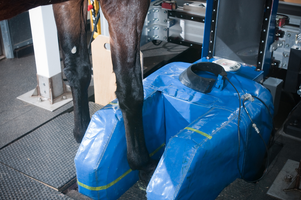
 Magnetic Resonance Imaging (MRI)
Magnetic resonance imaging (MRI) uses a powerful magnetic field, radio waves and a computer to produce detailed pictures of organs, soft tissues, and other internal body structures. The images can then be examined on a computer monitor or printed. MRI does not use ionizing radiation (x-rays), but detects the motion of hydrogen protons that are present in the body.
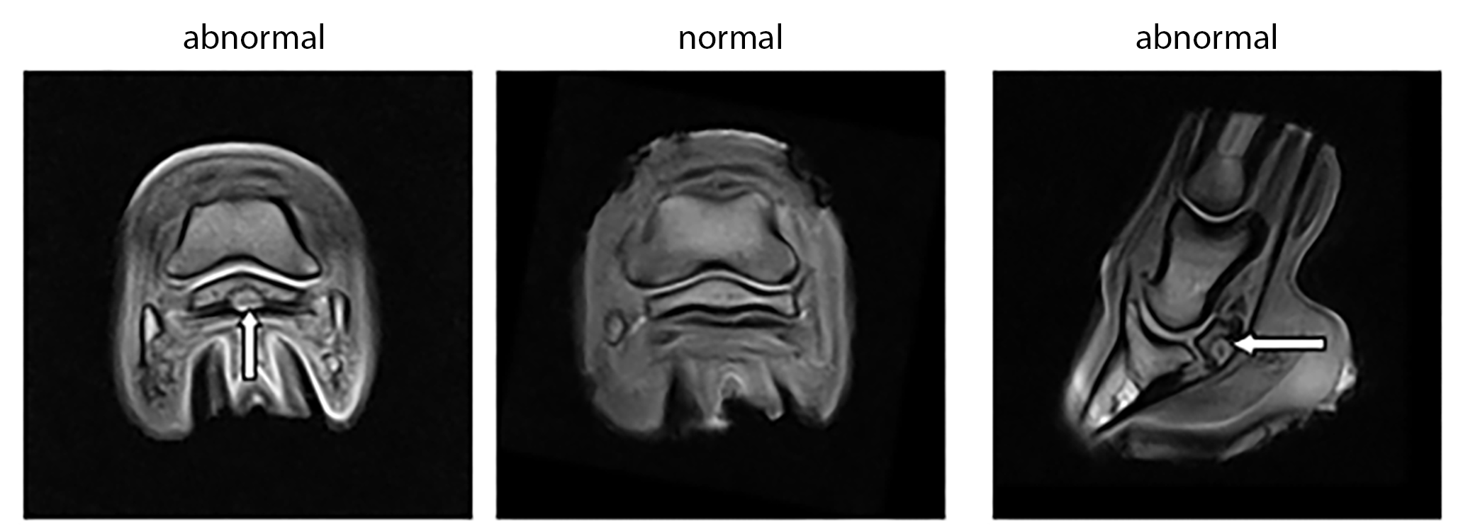
Detailed MR images allow veterinary radiologists to better evaluate parts of the body and certain diseases that may not be assessed adequately with other imaging methods such as x-ray, ultrasound or computed tomography. MR allows visualization of soft tissue structures not seen with x-ray, and is ideal for evaluating structures within the hoof that cannot be imaged using ultrasound. Identifies areas of inflammation, disrupted fiber pattern of tendons and ligaments, thickening of the joint capsule, and small bone lesions.