Positron Emission Tomography (PET)
In May of 2016, the UC Davis veterinary hospital acquired a positron emission tomography (PET) scanner, becoming the first veterinary facility in the world to utilize the imaging technology for equine patients. In association with the UC Davis School of Veterinary Medicine’s Center for Equine Health (CEH), the hospital launched the PET scanner in the summer of 2016. The unit had been acquired for research and clinical studies on lameness diagnosis in horses.
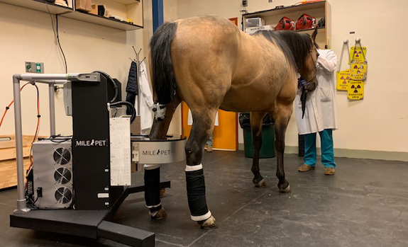
In 2015, veterinary radiologists from UC Davis were the first to ever image a horse using a prototype of the newly acquired scanner. This presented a major breakthrough in equine imaging, as PET imaging of horses had not been previously possible due to positioning restrictions of equine patients inside the standard PET instruments. With the new portable design, PET can now be applied to improve management of cases of equine lameness. The scanner weighs only 50 pounds, and was specifically designed for animal imaging.
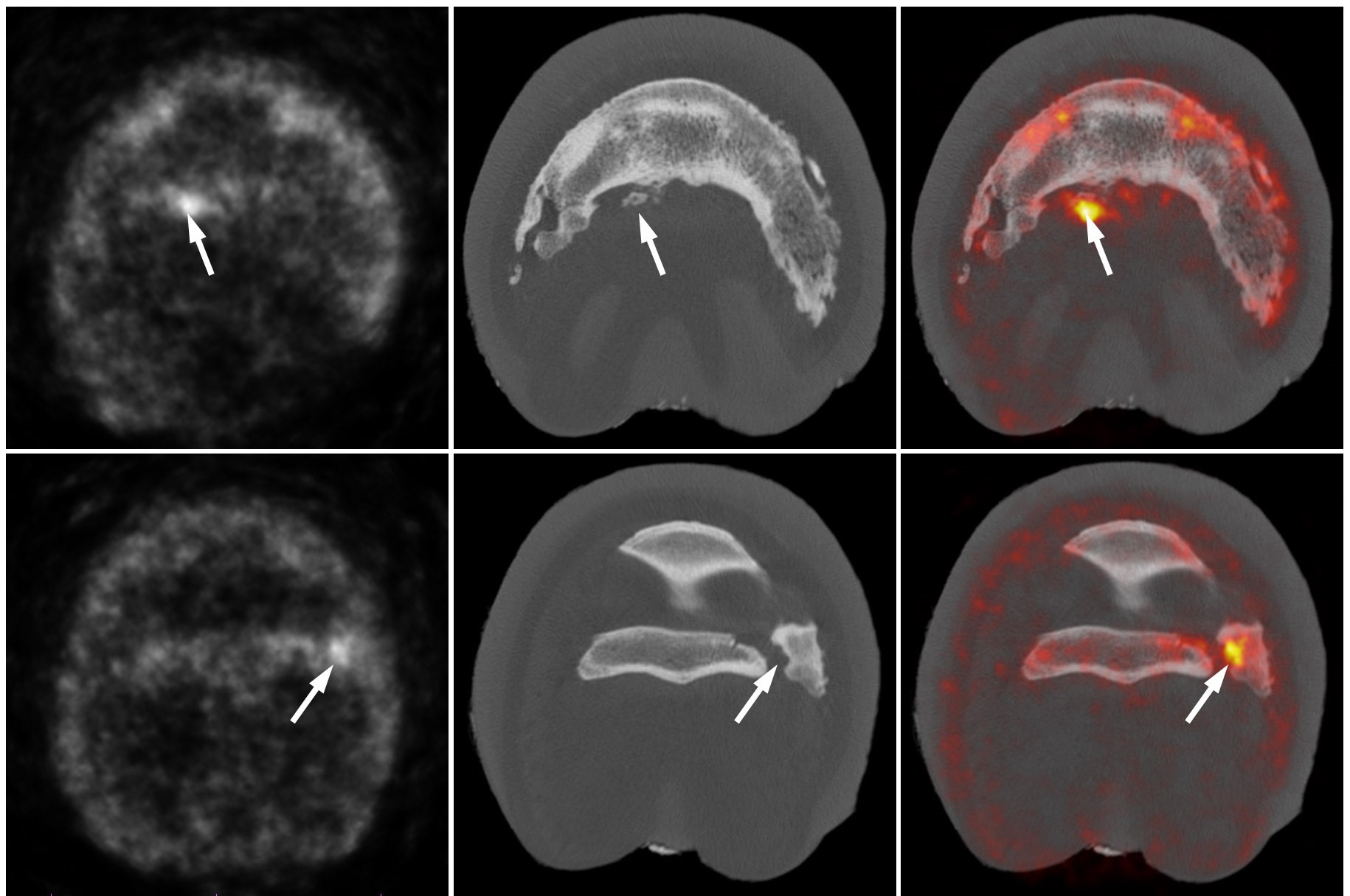
Although PET is mostly used in human medicine for the detection of cancer metastasis and for functional assessment of the brain, last year’s UC Davis study demonstrated promising applications for assessing the musculoskeletal systems of horses.
Equine standing positron emission tomography (PET) has been a game changer for monitoring racehorse health and guiding training and medical care, which has contributed to reduced injuries and fatalities. However, PET is not just for racehorses. With the rapid expansion in the availability of this technology, PET is becoming more accessible to diverse populations of horses. Standing PET can be used from the foot to the knee in the front limbs and the foot to the hock in the hind limbs.
Different imaging modalities can also be combined to provide the most accurate picture. In some cases, PET and X-rays will provide all the information necessary to decide on a treatment plan. In other cases, MRI of a specific area can be performed after PET, and the combination of PET and MRI data provides the most comprehensive assessment of the situation.