UC Davis radiologists were well represented at the 2022 American College of Veterinary Radiology (ACVR) annual scientific meeting, held recently in Reno, Nevada.
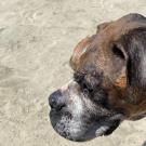
Bubbles, a 10-year-old male boxer, was brought to the UC Davis veterinary hospital’s Emergency Room following an inability to maintain his coordination accompanied with weakness in his hind limbs. Critical care specialists in the ER referred him to the Neurology/Neurosurgery Service for further evaluation. Following examination and an MRI, a tumor on Bubbles’ spinal cord was discovered.
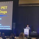
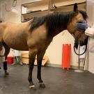
On August 5, UC Davis veterinary radiologist Dr. Mathieu Spriet presented "New Equine Imaging Options with Standing PET at UC Davis," an informative webinar to update veterinarians on the latest in positron emission tomography (PET) for horses.
UC Davis veterinary radiologist and pioneering researcher of equine PET scanning, Dr. Mathieu Spriet was recently selected as one of three veterinary specialists who will review all diagnostic imaging of horses competing in Australia’s 2021 Victorian Spring Racing Carnival.
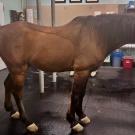
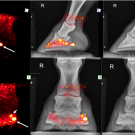
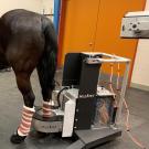
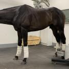
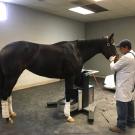
Standing equine positron emission tomography (PET) imaging is not just for racehorses anymore. In the first four months since the installation of the MILEPET scanner at the UC Davis veterinary hospital, 100 horses have been imaged; more than half were performance and pleasure horses.
The UC Davis standing equine positron emission tomography (PET) scanner is officially in use at Golden Gate Fields racetrack in Berkeley, CA, providing imaging at the molecular level to monitor racehorse health and guide training and medical care.
On April 14, 2021, local television morning show "Good Day Sacramento" visited the UC Davis Center for Equine Health. Dr. Mathieu Spriet showcased the latest in equine PET scanning technology.
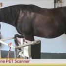
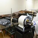
The equine Positron Emission Tomography (PET) scanner pioneered by the UC Davis School of Veterinary Medicine, in collaboration with LONGMILE Veterinary Imaging, is now in heavy use at Santa Anita Park in Southern California. In just over six months since the installation in December 2019, with the financial support from the Stronach Group, more than 100 scans have been performed with the “MILEPET” (Molecular Imaging of Limbs in Equids), the PET scanner specifically designed to acquire images on horses without the need to lay them down.
Dr. Mathieu Spriet, an associate professor in the Department of Surgical and Radiological Sciences, recently passed boarding examinations to become a founding member of the American College of Veterinary Radiology’s (ACVR) new subspecialty of Equine Diagnostic Imaging.